clinicians have a wide array of radiologic modalities at their disposal to aid them in noninvasive diagnosis. Despite the introduction of highly specialized imaging modalities, radiologic test such as Chest radiographs and ultrasound Continue to serve a vital role in the diagnostic approach to pt care. Diagnostic imaging in internal Medicine
at most institutions, CT is available on an emergent basis and is invaluable for initial evaluation if pts with trauma, suspected CNS hemorrhage, or ischemic stoke. MRI and related techniques (MR angiography, functional MRI, MR spectroscopy) provided remarkable resolution of many tissues including the brain, vascular system, joints, and most large organs. Radionuclide scans including positron emission tomography (PET)can provided functional assessment of organs or specific regions within organs, combination of PET with MRI or CT scanning provides highly informative images of the location and configuration of metabolically active lesions such as cancer. increasingly, internists are being trained in the use of ultasound to assist with line placement, thyroid nodules, cardic sounds and abdominal abnormalities.
Chest Radiography Diagnostic imaging in internal Medicine
Can be obtained quickly and should be part of the standard evaluation for pts with cardiopulmonary complaints.
Is able to identify life-threatening conditions such as pneumothorax, intraperitoneal air, pulmonary edema, pneumonia, and aortic dissection.
Is most often normal in a pt with an acute pulmonary embolus.
Should be repeated in 4-6 weeks in apt with an acute pneumonic process to document resolution of the radiographic infiltrate.
Is used in conjunction with the physical examination to support the diagnosis of congestive heart failure, Radiographic findings supporting the diagnosis of heart failure include cardiomegaly, cephalization, kerley b lines, and pleural effusions.
Should be repeated frequently in intubated pts to examine endotracheal tub position and the possibility of barotrauma.
Helps you identify alveolar or airspace disease. Radiographic features of such diseases include inhomogeneous, patchy opacities and air- Broncho grams.
Helps to document the free flowing nature of pleural effusions, Decubitus views should be obtained to exclude loculated pleural fluid prior to attempts to extract such fluid.
Abdominal Radiography Diagnostic imaging in internal Medicine
Should be the initial imaging modality in a pt with suspected bowel obstruction. Signs of small-bowel obstruction on plain radiographs include multiple air-fluid levels, absence of colonic distention, and a “stepladder” appearance of small-bowel loops.
Should not be perfomed with barium enhancement when perforated bowel, portal venous gas, or toxic megacolon is suspected.
Is used to evaluate the size of bowel:
- Normal small bowel is <3 cm in diameter.
- Normal caliber of the cecum is up to 9cm with the rest of the large bowel up to 6 cm in diameter.

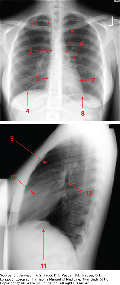
Normal chest radiograph-review of anatomy. 1. Trachea. 2. Carina 3. Right atrium. 4. Right hemi diaphragm 5. aortic knob 6. Left hilum 7. Left ventricle. 8 left hemi diaphragm (with stomach bubble). 9. Retrosternal clear space. 10. Right ventricle. 11. Left hemi diaphragm (with stomach bubble). 12. Left upper lobe bronchus.
Ultrasound Diagnostic imaging in internal Medicine
1. is more sensitive And specific than CT scanning in evaluating for the presence of gallstone disease.
2. Can be used To assist with central line placement and with peripheral access when challenging.
3. can readily identify the size of the kidneys in a pt with renal insufficiency and can exclude the presence of hydronephrosis.
can expeditiously evaluate for the presence of peritoneal fluid in a pt with blunt abdominal trauma.
is used in conjunction with Doppler studies to evaluate for the presence of arterial atherosclerotic disease.
Should be used to localize loculated pleural and peritoneal fluid prior to draining such fluid.
Can determine the size of thyroid nodules and guide fine-needle aspiration biopsy.
Can determine the size and location of enlarged lymph nodes, especially in superficial locations such as in the neck.
Is the modality of choice for assessing known or suspected scrotal pathology.
Should be the first imagining modality utilizer when evaluation the ovaries.
Most Related Articales on
Top 10 Health issues 2024


protogel protogel protogel
Hello it’s me, I am also visiting this site
regularly, this web site is in fact nice and the users are in fact sharing
nice thoughts.
Thanks for another informative website. Where else may I get that kind of
info written in such an ideal means? I have a venture that I’m simply now operating on, and I
have been on the look out for such info.
For most recent news you have to pay a visit world wide web and on internet I found this web site as a best site for most recent updates.
Hey there I am so happy I found your webpage, I really found
you by accident, while I was browsing on Yahoo for something else, Anyways I am
here now and would just like to say many thanks for a tremendous post and a all round enjoyable blog (I also love the theme/design), I don’t have
time to browse it all at the moment but I have book-marked it
and also added in your RSS feeds, so when I have time I will be back to read a lot more, Please do keep up the superb work.
We stumbled over here from a different website and thought
I may as well check things out. I like what I see so now i am following you.
Look forward to finding out about your web page yet again.
After looking at a few of the blog posts on your web site, I honestly
like your way of writing a blog. I saved as a favorite it to my bookmark webpage list and will be checking back in the near future.
Take a look at my web site too and tell me what you think.